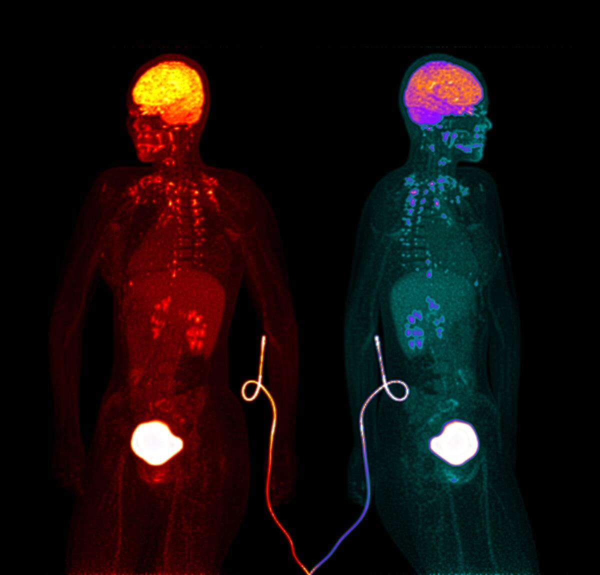
Metabolic Symphony
This image captures the hidden music of the human body —
glucose flowing, organs glowing, life unfolding in light.
A radioactive tracer travels through the veins,
revealing where energy is consumed,
where thought ignites and silence rests.
The brain shines brightest —
a beacon of constant hunger for sugar and meaning.
The mirrored figures are the same person,
seen through two spectral lenses:
one in warm fire, one in cool night.
Together they show how the invisible becomes visible —
how life itself can be traced by light.
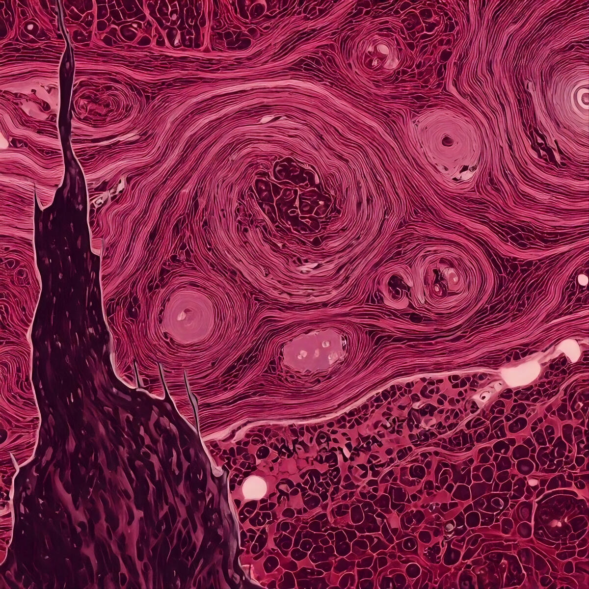
Tissue Turbulence
The starry night, a painting that at first glance depicts a serene village under a clear night sky, but upon looking closely we understand Van Gogh's turbulent perception of the world.
Similarly, tissues can appear deceptively normal on whole-slide imaging. Only when we zoom in do the hidden details emerge, interstitial fat deposition, narrowed or damaged vessels, inflammatory infiltrates, cellular atrophy, and other subtle changes that reflect underlying biological processes.
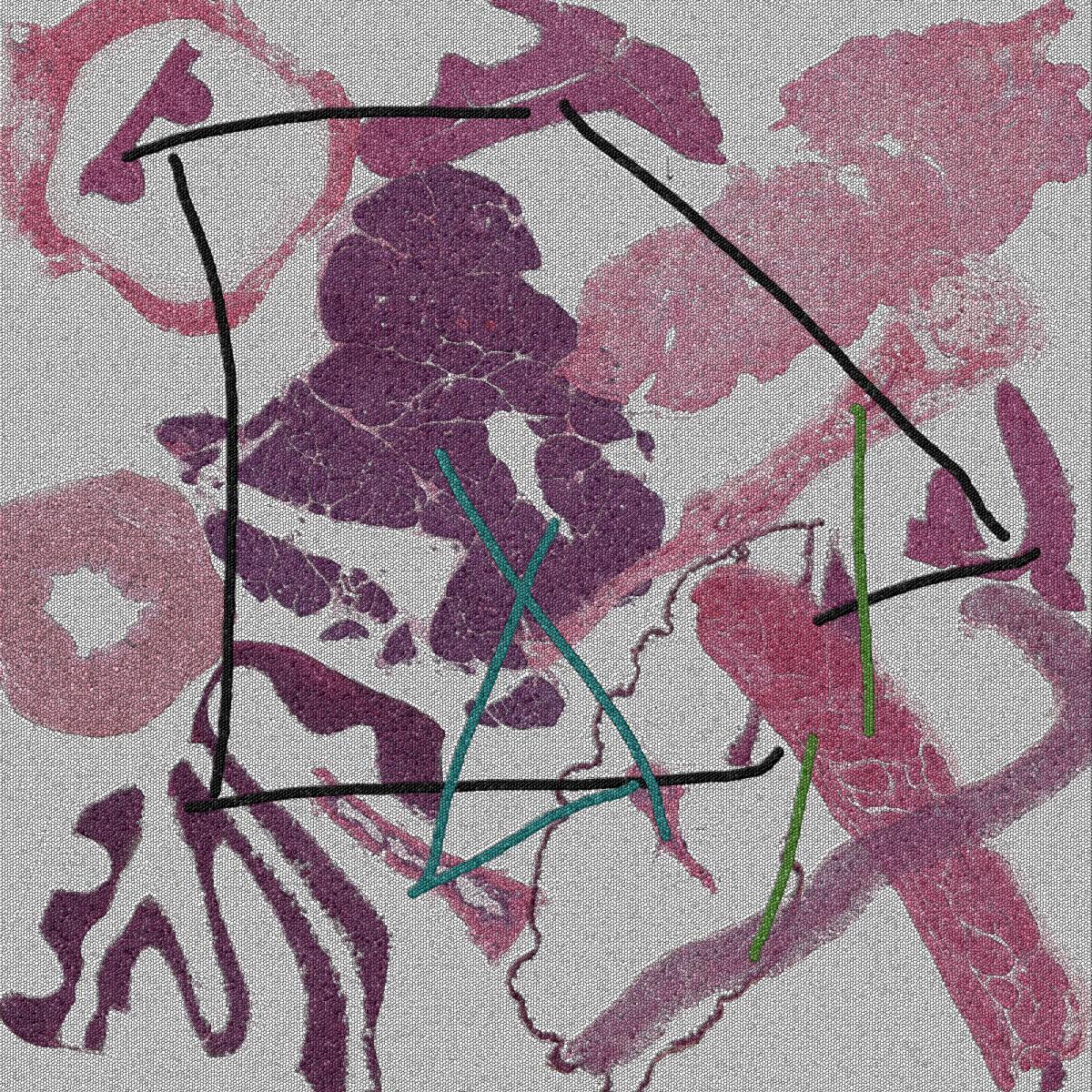
The puzzle of microanatomy
What are we made of? Is it the atoms in our body, the cells in our brain?
At the mesoscale (hundreds of microns to centimeter scale) our cells arrange themselves into microanatomical structures each associated with a function for the respective organ - the mucosa of the stomach provides a barrier, the muscle movement for digestion; pancreatic ducts drive digestive juices out of the pancreas; the outer layer of the coronary artery provides support and elasticity.
Finding these microanatomical domains is not easy, but that's one of our projects. In this Frame, you get to participate! What organs are represented? Which domains are connected? What is their coordinated function?
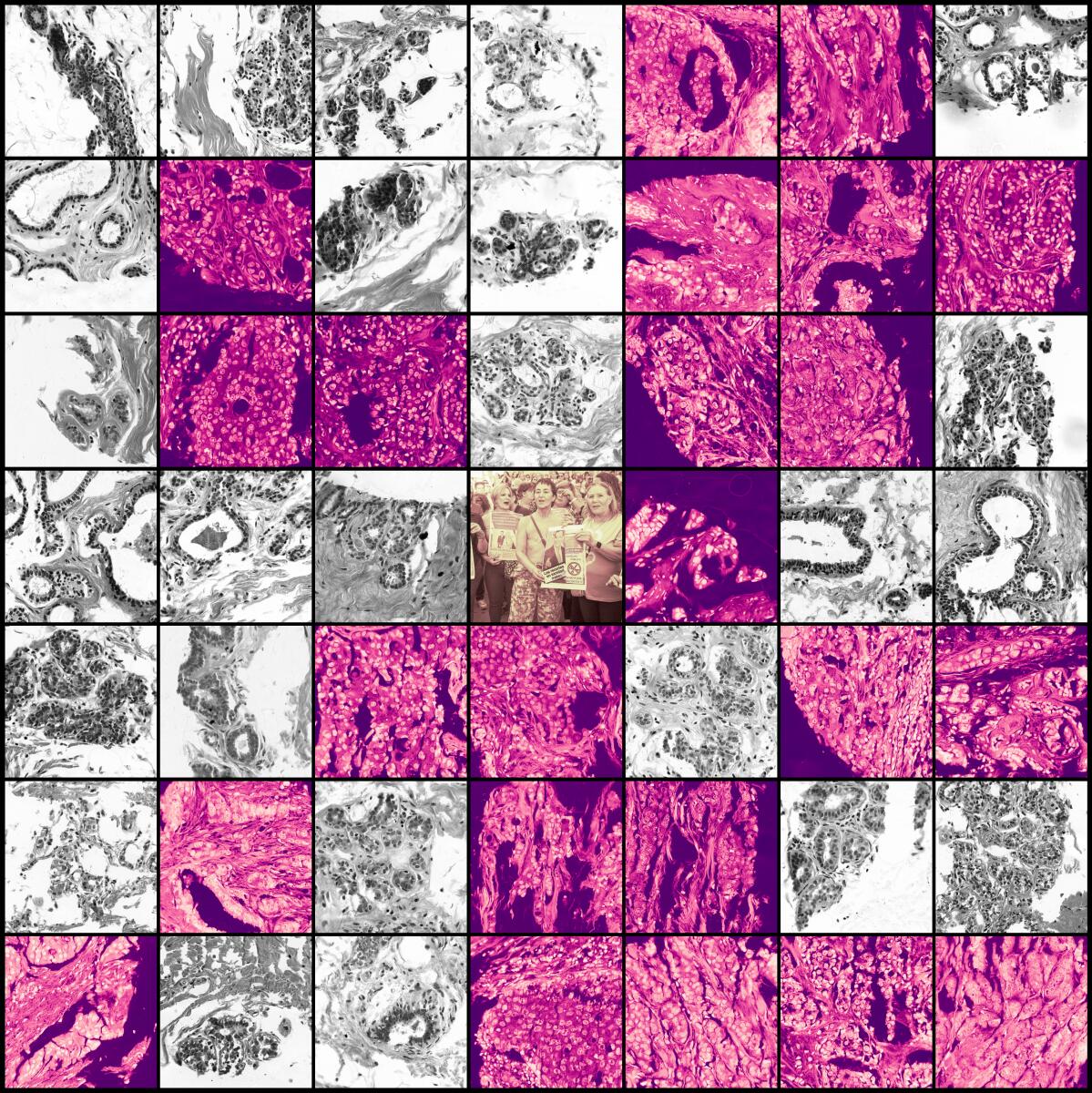
Faces behind the data
Histopathology is a cornerstone of breast cancer diagnosis, revealing under the microscope the difference between benign (grey) and malignant cells (magenta). Before reaching this stage, patients rely on screening methods such as mammography to flag potential disease. Amid the influx of medical data, it is easy to lose sight of the individuals behind the numbers, the patients who must remain the focus of scientists, clinicians, and administrators alike. In October 2025, more than 2,300 women in Andalusia, Spain, learned that they had not been informed of abnormal mammogram results due to failures in the regional breast-cancer screening system, sparking public outrage, protests, and an official investigation.
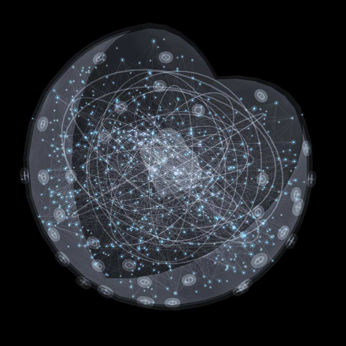
Nuclear Constellation
For centuries, humans looked to the sky, mapping stars and constellations in search of meaning and predictions about the future. Today, we look inward: within the nucleus of every cell lies a dense network of interacting proteins – the molecular machinery that regulates and maintains life. This visualization mirrors the nuclear cosmos within us, a constellation of molecular interactions shaping the architecture of cellular regulation and communication.
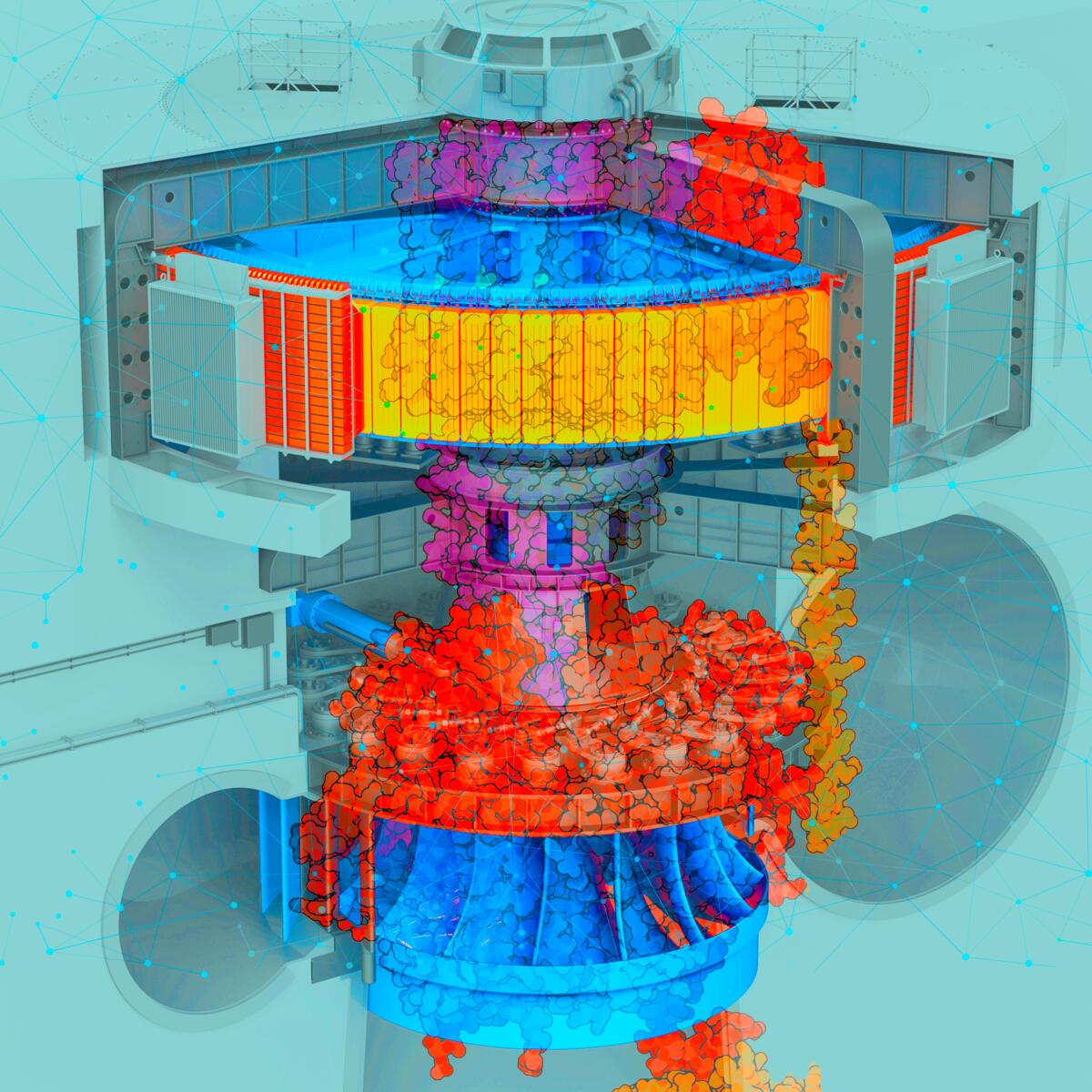
Proteins - Life's Molecular Engines
This artwork depicts the ATP synthase complex as a hydroelectric turbine, drawing a parallel between engineered systems and molecular machinery. Just as a dam harnesses water flow to generate electricity, ATP synthase channels a proton gradient like a "molecular motor" to produce ATP, the chemical energy currency for all cellular processes. This piece reflects the LBI for Network Medicine's core mission: to understand life as a complex system of molecular interactions and to decipher the mechanistic basis of health and disease.
DNA Bases
Is it the structure?
This piece reimagines the famous DNA "X" as pure text art: the image is rebuilt entirely from the four DNA letters (A, C, G, T), lightly color-tinted by base and given a gentle, diagonal depth-of-field so your eye settles on the central cross. The underlying frame references Photograph 51—the 1952 X-ray diffraction image of DNA made at King's College London by Rosalind Franklin and her student Raymond Gosling—which revealed the telltale "X" pattern that helped lead to the double-helix model (Watson & Crick, 1953). In other words: a pivotal scientific snapshot is translated into the alphabet of life itself, turning data into typography while nodding to the moment DNA's structure came into focus.